Eohumeralcemic refers to a specific metabolic condition that affects bone and joint function. Clinicians identify eohumeralcemic by elevated humeral calcium levels and by related systemic signs. This article defines eohumeralcemic, lists common symptoms, explains causes, and outlines diagnosis and treatment in clear terms. It gives actionable steps that clinicians and patients can use in 2026.
Table of Contents
ToggleKey Takeaways
- Eohumeralcemic is a metabolic condition characterized by elevated calcium levels near the humerus, leading to bone and joint issues primarily around the shoulder.
- Early symptoms include mild shoulder pain and stiffness, while advanced cases cause constant pain, loss of function, and possible deformity near the humerus.
- Primary eohumeralcemic often results from parathyroid disorders, while secondary cases stem from conditions like chronic kidney disease or vitamin D imbalances.
- Diagnosis involves serum calcium tests, bone markers, and imaging techniques such as X-rays, CT, MRI, and DEXA scans to differentiate from similar disorders.
- Treatment targets the root cause, including surgery for parathyroid adenomas, mineral balance correction in renal cases, and medications like bisphosphonates to reduce bone loss.
- Long-term management includes rehabilitation for shoulder function, lifestyle guidance on calcium and hydration, and regular monitoring through labs and imaging to prevent complications.
What Is Eohumeralcemic? A Clear Definition And Clinical Overview
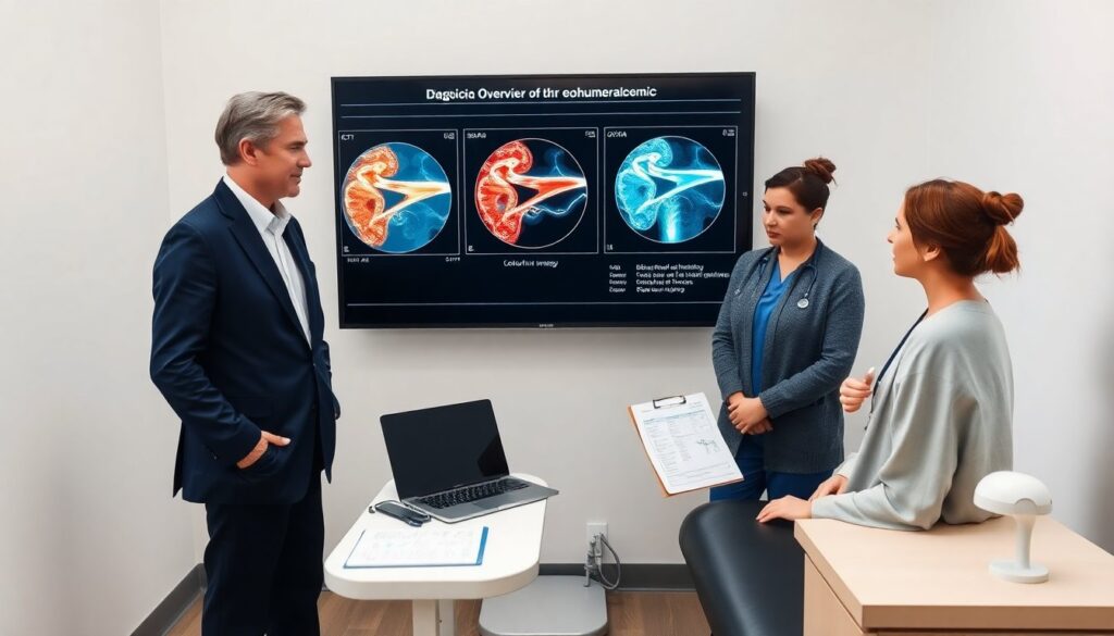
Eohumeralcemic describes a condition in which calcium concentrates abnormally near the humerus and in related tissues. Doctors measure serum calcium and local bone markers to confirm eohumeralcemic. Patients with eohumeralcemic often show changes in bone density near the shoulder and in nearby soft tissue. Clinicians classify eohumeralcemic as primary when a parathyroid or bone disorder drives it. They call it secondary when another disease, like renal failure or vitamin D disorder, drives it. Researchers use the term to group cases with similar laboratory and imaging patterns.
Common Symptoms And Signs To Watch For
Patients with eohumeralcemic report shoulder pain and reduced range of motion. They may feel stiffness and weakness in one or both arms. Physical exam often shows localized tenderness over the humerus. Lab tests may show high serum calcium and altered bone turnover markers. Imaging may show cortical thickening, focal osteolysis, or calcified soft tissue near the shoulder. Systemic signs can include fatigue, nausea, and thirst when serum calcium rises. Clinicians should track symptom onset, duration, and any recent illness or medication change that might link to eohumeralcemic.
Early Versus Advanced Symptom Patterns
Early eohumeralcemic presents with mild pain and intermittent stiffness. Patients in early stages often keep normal daily function. Advanced eohumeralcemic shows constant pain and marked loss of shoulder function. Advanced cases can show visible deformity or recurrent fractures near the humerus. Lab values often show higher calcium and more disrupted bone markers in advanced cases. Imaging moves from subtle cortical changes in early cases to clear bone loss or large calcifications in advanced cases. Early diagnosis improves chances to slow progression of eohumeralcemic.
Causes And Risk Factors Behind Eohumeralcemic
Parathyroid adenoma and hyperparathyroidism cause many primary eohumeralcemic cases. Chronic kidney disease and prolonged immobilization cause secondary cases. Excess vitamin D intake can cause calcium shifts that look like eohumeralcemic. Age and osteoporosis raise risk for bony changes near the humerus. Certain cancers that affect bone can trigger localized calcium changes that mimic eohumeralcemic. Use of thiazide diuretics or lithium can raise serum calcium and contribute to eohumeralcemic. Clinicians should assess medications, endocrine history, and renal function when they evaluate possible eohumeralcemic.
How Eohumeralcemic Is Diagnosed: Tests, Imaging, And Differential Diagnosis
Doctors diagnose eohumeralcemic with labs and targeted imaging. They order serum calcium, parathyroid hormone, vitamin D, and renal panels. They add bone turnover markers when they need detail. X-rays detect cortical changes and calcifications. CT and MRI show soft tissue and subtle bone involvement. Dual-energy X-ray absorptiometry (DEXA) helps measure bone density near the shoulder. Biopsy rarely proves necessary but it rules out malignancy when imaging looks unclear. Doctors distinguish eohumeralcemic from osteoarthritis, rotator cuff disease, metastatic bone disease, and infection by combining lab and imaging results.
Treatment Options And Ongoing Management Strategies
Treatment for eohumeralcemic targets the underlying cause and the local bone changes. For parathyroid-driven cases, surgery to remove an adenoma often lowers serum calcium and helps eohumeralcemic. For renal-related cases, correcting mineral balance and using phosphate binders helps control calcium and limits eohumeralcemic. Clinicians use bisphosphonates or denosumab to reduce bone resorption when bone loss drives eohumeralcemic. Pain control may include acetaminophen, short courses of NSAIDs, or targeted steroid injections for inflammatory flares. Follow-up labs and periodic imaging let clinicians track response to treatment and adjust care for eohumeralcemic.
Lifestyle, Rehabilitation, And Monitoring For Long-Term Outcomes
Patients with eohumeralcemic benefit from structured rehabilitation to restore shoulder function. Physical therapists design progressive range-of-motion and strengthening programs that address deficits from eohumeralcemic. Patients should maintain adequate hydration and follow dietary calcium and vitamin D guidance set by clinicians. Clinicians schedule serum calcium checks every few weeks after major treatment and then every three to six months when stable. Repeat imaging at three to twelve months helps confirm healing of local bone changes from eohumeralcemic. Early activity modification and close follow-up lower the risk of fractures and persistent disability from eohumeralcemic.